The experience of Hospital Universitario La Paz and 3D printing
The Hospital Universitario La Paz was built more than 50 years ago as a european reference center in various medical and surgical specialties. The promotion of research and teaching have marked its performance over the years, always at the service of the patient and seeking healthcare excellence.
In 2022, in their constant search for excellence and innovation, Hospital La Paz contacted 3DZ to evaluate the possibilities of different 3D technologies in the development of models for reconstructive surgery.
The challenge: to create a manufacturing protocol for customized, 3D printed and sterilizable ear models for autogenous ear reconstruction
Auricular reconstruction in patients with microtia is a constant challenge for the surgeon, being considered one of the most technically difficult and demanding procedures.
The main reconstructive option, since the 1920s, has been reconstruction using costal cartilage. However, due to the extremely complex and unique anatomy of the ear, this procedure requires a steep learning curve, which is highly dependent on the surgeon’s artistry and technical skills.
Traditionally, they have used a 2D radiographic film template, taken from the contralateral ear. The main difficulty is to transfer the 3D characteristics of the anatomical elements of the ear (thickness, height, depth, reliefs).
The challenge was to find a 3D model of the contralateral ear that could be taken quickly, as these are often children, who are mobile or uncooperative.
Doctor of the Oral and Maxillofacial Surgery Service at the Hospital Universitario La Paz
Dr. Juan Pablo Rodríguez-Arias:
“For years, we had already been taking a model of the contralateral ear with a plaster cast, but this could not be sterilized or used in the operating room as a guide. Besides, obtaining this model is very cumbersome and uncomfortable for children,” says Dr. Rodríguez Arias, who together with Dr. Mercedes Martín, Dr. Elena Gómez and Dr. Jesús Manuel Muñoz, make up the Children’s Maxillofacial Surgery Department headed by Dr. José Luis Cebrián. “In other hospitals, they were starting to take the CT contralateral ear model, but we wanted to avoid radiation. On the other hand, we thought it would make the surgery even easier, to have the 3D models of the pieces that we subsequently carved to reconstruct the ear, and not the whole ear.”
So it was that in early 2022 the Maxillofacial Surgery department contacted 3DZ to acquire a 3D scanner.
The objectives
1
Obtain accurate ear shape and detail, quickly and accurately.
2
Obtain segmented models of the ear, sterilizable.
3
To increase the comfort of the patient, usually children who are mobile or uncooperative.
4
Do not add comorbidity or radiation in the pre-intervention phase.
5
To facilitate the surgeon's work by reducing surgical time.
The solution
The role of Alessandro Gutierrez Venturini, an engineer at the hospital’s 3D Management Laboratory, is particularly relevant here. Based on close surgeon-engineer communication, the Maxillofacial Surgery service and the 3D Management Laboratory developed a protocol for the acquisition, processing and digital segmentation of the ear images, in order to obtain the desired ear models.
“It should be noted that this protocol is technically demanding, requires expertise in 3D design and therefore also a considerable learning curve, especially during the preoperative planning phase. We consider it essential that in this phase, engineer and surgeon work together to contribute our technical and clinical vision and experience,” says Alessandro Gutiérrez before detailing the process.
After the consultancy with 3DZ, the hospital acquires an Artec Eva 3D scannerand receives training from our highly qualified technical staff to get the most out of the use of 3DZ equipment.
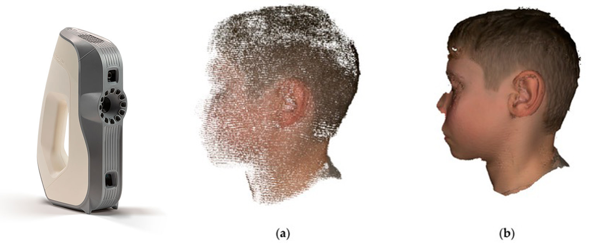
1 - Atrial surface scanning with Artec EVA
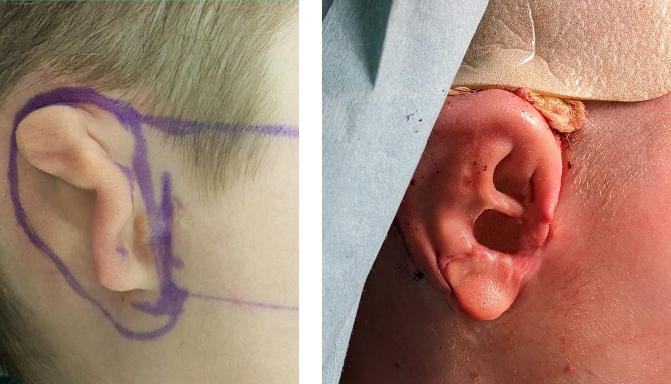
The first step of the protocol consists of acquiring images of the subject’s healthy ear. In this case an 8 year old male with lobar type microtia on the right side. High accuracy is needed to capture the detailed features of the auricular structure, which are essential for a successful reconstruction.
In this case, the Artec Eva structured light surface 3D scanner was used, which allows the capture of surfaces with a 3D resolution of up to 0.2 mm, at a reconstruction rate of 16 fps, while also saving the texture of the model. The average image acquisition time in this case was 65 s.
The obtained point cloud data were processed in the native Artec Studio 16 Professional software. After a process of discarding, registration, analysis, alignment, noise removal, a fusion of all frames into a single mesh is performed.
In image (a) we see the first data captured from the point cloud before post-processing; in (b) the final reconstruction of the 3D digital model after post-processing.
For the next stage, the obtained mesh was exported as an STL file.
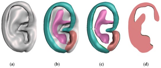
2 - Preparation of digital models
Among other steps, the exported mesh was processed with the modeling software in order to design the new components of the patient’s affected ear.
The models shown in the image were exported in STL format for the 3D printing phase:
(a) refined and mirrored healthy ear; (b) segmentation of the helix (blue), antihelix (purple) and tragus (brown); (c) sculpted anatomical parts; (d) base frame model.
This is the modeling of the ear and anatomical parts for surgical planning.
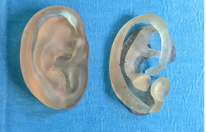
In the photo, 3D printed models of the ear (left) and the different anatomical parts (right).
After printing, the models were removed from the build platform and washed for 20 minutes in a Form Wash (Formlabs) with 99% isopropyl alcohol to clean the parts and remove liquid resin. They were then post-cured at 60 °C for 30 min in a Form Cure (Formlabs) to achieve biocompatibility and optimal mechanical properties.
Finally, the models were sterilized and used in the operating room.
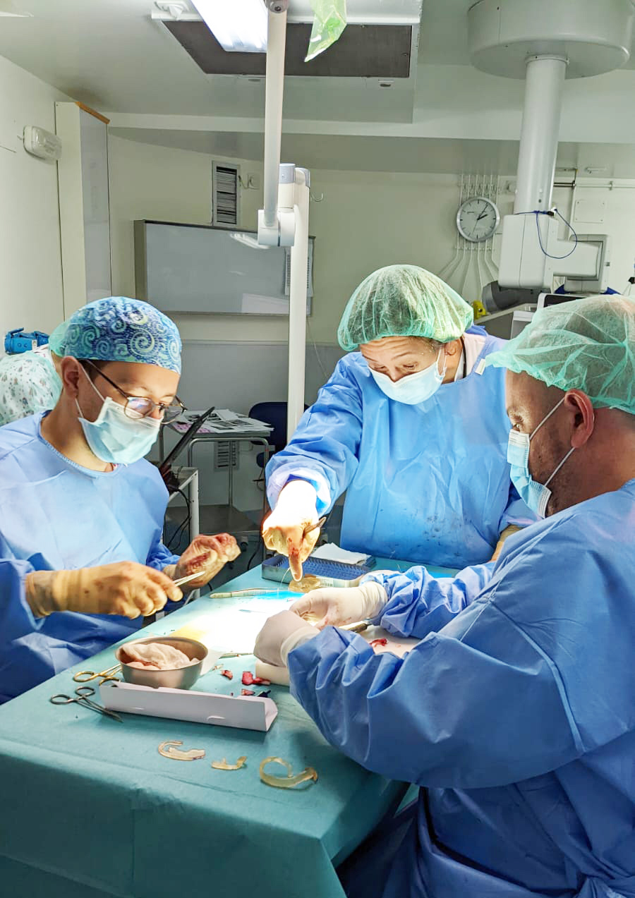
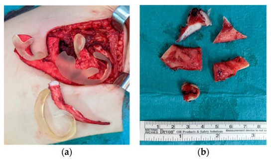
4 - Structural fabrication
The size of the normal pinna and the type of microtia determine how many cartilage fragments are removed.
Thanks to these 3D models, several cartilage fragments were left over and repositioned in the costal pocket so that they could be used as Firmin P1 in the second phase of the procedure.
RESULTS
Thanks to the patient-specific sterilized 3D models, the surgeon was able to hold the grafts, rotate them and analyze their shapes and relief. Preoperative planning of the surgery during joint digital processing with the 3D Management Laboratory engineer saved both surgical time and the amount of cartilage, without losing the artistic element inherent to this type of surgery.
Key benefits of using 3D technologies in reconstructive surgery
The precision of the 3D models printed in biomedical resin makes it easier to handle them safely and without fear of contamination of the biological material. “Having the sterile models on the operating room table is a differential factor, since it allows us to directly compare 1 to 1, piece by piece, the costal components that make up our auricular reconstruction“, says Dr. Rodriguez Arias.
This shortens surgical time considerably, which is beneficial forboth the patient and the medical team. Less time in the operating room reduces the risk of complications and improves patient recovery, resulting in cost savings.
With precise 3D models, only the amount of cartilage necessary for the intervention is removed, avoiding the unnecessary removal that used to occur in the two-dimensional method. “This not only reduces the child’s postoperative pain, but also decreases recovery time and the risk of complications such as pneumothorax,” says Rodriguez-Arias. “In addition, our protocol allows the reconstruction of any subtype of microtia, whether total or partial, further optimizing the removal of the rib cartilage”.
The fabrication of the model in biomedical resin provides a safer and more sterile working environment. “It is very comfortable to manipulate both the models and the cartilage on the same operating table, without the need to change gloves…, without fear of contamination of the field”.
Three-dimensional templates have been shown to reduce surgery time, saving hospital resources that could offset the main cost of the printer and the type of scanner chosen. “The decrease in surgical time is a great saving, since it is estimated that the cost per minute in the operating room varies between €30 and €50. Although we have not completed the study, we estimate that we have reduced surgical time by one hour“, assures Dr. Rodriguez-Arias.
The future
“We want to continue optimizing our protocol, as well as extrapolating and perfecting it in other interventions in such a complex body area as the maxillofacial, of great three-dimensional complexity, and with both aesthetic and functional components and demands (…). ) On the other hand, we have open studies to evaluate the use of 3D technologies in the postoperative follow-up in craniosynostosis surgery, which we carry out jointly with the Department of Child Neurosurgery, and to be able to optimize our surgical technique according to these findings“, concludes Dr. Juan Pablo Rodriguez-Arias.
3DZ, an expert partner
At 3DZ we are distributors of the best 3D printing and scanning brands with more than a decade of experience. We accompany companies in choosing the best 3D technology for their specific needs.
Contact us, our team is at your disposal.